


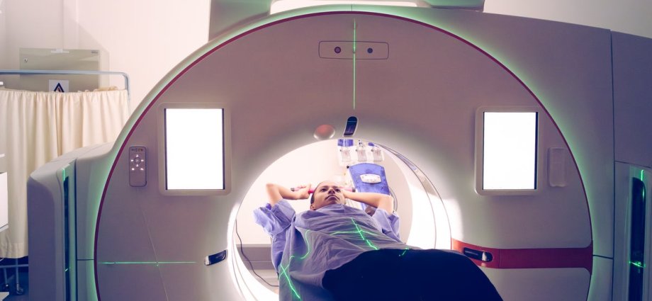
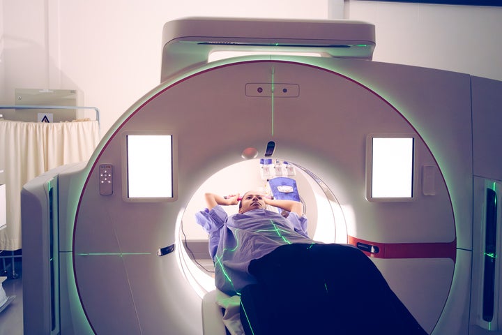
A new artificial intelligence model can detect signs of pancreatic cancer on CT scans up to three years before a clinical diagnosis, according to a study.
The technology, developed by researchers at the Mayo Clinic, identifies “subvisual” patterns in the pancreas that are invisible to the human eye, the study published in the journal Gut found.
By the time most patients develop symptoms like abdominal pain or weight loss, the cancer has typically reached an advanced stage. Pancreatic cancer currently has a five-year survival rate of just 13 percent.
The AI framework, called Radiomics-based Early Detection MODel, was trained using nearly 1,000 CT scans from patients who were originally screened for unrelated conditions but later developed pancreatic cancer.
In a head-to-head comparison with board-certified radiologists, the AI was significantly more effective at spotting early markers of the disease.
Pancreatic tumors are notoriously difficult to find because the organ is located deep within the abdomen, making physical exams ineffective. There is also no routine screening available for the general public.
On an independent test set of 493 scans, the model identified occult cancer with a 73 percent sensitivity rate — nearly double the 39 percent achieved by human doctors. For scans taken more than two years before a formal diagnosis, the AI was nearly three times as sensitive as the radiologists.
“We knew, based on the biology of the disease, that this is not something which is coming all of a sudden in three months,” Dr. Ajit Goenka, a Mayo Clinic radiologist and study author, said. “We knew that the signal was there. We just needed to find a way to be able to detect it.”
The AI model works by analyzing “radiomic” features — minute textural disruptions in the organ’s tissue. Goenka said that the model could identify abnormal cells that protect cancer from the immune system, a signature scientists have long recognized but struggled to visualize on standard imaging.
Dr. Daniel Jeong, a radiologist at Moffitt Cancer Center who was not involved in the research, spoke about the limitations of current manual reviews.
“I analyze these images every day,” Jeong told NBC News. “We’re really looking for a measurable mass that could represent the cancer. So these tumors need to grow to a certain level to become visible.”
The research marks a significant milestone, but experts caution that the tool is not yet ready for widespread use. The model is currently being evaluated in a clinical trial that requires three to five years of patient monitoring to confirm its accuracy in real-time.
Researchers suggest the technology could eventually serve as a triage tool for high-risk individuals, such as those with a family history of the disease or new-onset diabetes.
“In a disease where we have been just wandering in darkness for decades, this is a milestone that shows us the finish line, but we still have to get to the finish line,” Goenka said.
The study joins several recent advancements in the field, including early-stage trials for mRNA vaccines and experimental drugs that have shown promise in extending life expectancy for those with advanced cases.