










Want to bookmark your favourite articles and stories to read or reference later? Start your Independent Membership today.
Join today
Already a member?
Log in
Scientists have uncovered how extreme or frightening moments trigger a rapid biological chain reaction in the brain that forces it to lock in lasting memories, while letting everyday routines fade away.
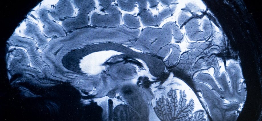
Researchers at the University of Utah found that a small, almond-shaped structure deep inside the brain – known as the amygdala – acts as a vital gatekeeper during moments of sudden shock, danger, or excitement.
The review, published in the journal Frontiers for Young Minds, brings together decades of laboratory research and human clinical trials to map how the brain filters human experience.
Normally, the brain discards the vast majority of mundane daily routines – such as a routine trip to the shops – to avoid becoming overwhelmed by trivial details.
But when something intense or deeply emotional happens, the amygdala switches on and prompts neighbouring brain regions to preserve the moment for long-term recall.

The chain reaction begins the moment a person encounters a sudden threat or intense surprise.
Adrenal glands sitting atop the kidneys release adrenaline into the bloodstream, stimulating a region in the brainstem.
That brainstem region then delivers a chemical messenger called norepinephrine directly into the amygdala.
Once activated, the amygdala does not act as a storage vault itself.
Instead, it functions as an internal alert hub that directs the hippocampus—a seahorse-shaped structure essential for memory formation and retrieval—to record sensory details as an event unfolds.
By sending electrical signals and protein-triggering messages to the hippocampus, the amygdala strengthens connections between brain cells.
This prompts the hippocampus to gather sights, sounds, smells, and physical sensations from separate neural hubs, including the visual, auditory, and olfactory cortices, binding them together for later recall.

To test this circuit directly in humans, researchers studied epilepsy patients who already had electrodes surgically implanted in their brains for medical monitoring.
During trials, participants were shown a series of images on a computer screen before doctors delivered a tiny, imperceptible electrical pulse directly to the amygdala.
When tested the following day, patients were significantly better at recognising the pictures paired with the electrical stimulation than those viewed without it.
By contrast, routine physical habits – such as tying shoelaces or kicking a ball – bypass this emotional alert system entirely, relying instead on a brain region called the striatum.
The researchers stressed that human recall does not work like replaying an unedited film.
When retrieving a past event, the brain stitches stored sensory fragments back together, occasionally filling in missing gaps based on surrounding context.
Mapping how this circuit operates marks a major step forward in understanding human memory, opening the door to targeted therapies for severe memory loss, dementia, and trauma-related conditions.