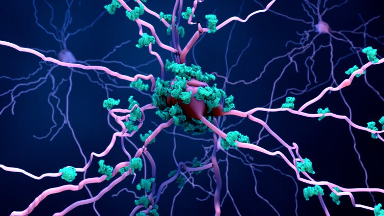
Alzheimer’s disease is a devastating condition that is currently unstoppable and incurable. The main cause of the disease is the loss of neurons and other brain cells in the brain – also know as degeneration. This degeneration is what leads to problems with memory and other cognitive functions.
Researchers can tell which neurons die first or exhibit increased vulnerability to Alzheimer’s disease based on where they’re located in the brain and what they look like. But they don’t know what genes or proteins these neurons express. Knowing these factors is important for recognising and identifying the changes in specific cells that happen when disease is present.
Now, a recent study has shown that neurons expressing a specific protein are more vulnerable to degeneration. Understanding which neurons are more vulnerable – and why – might allow researchers to develop targets for potential treatments in the future.
To conduct their study, scientists performed a post-mortem brain analysis on people who had Alzheimer’s disease. To see how far the disease had progressed, they began by looking for build-ups of the protein tau in different parts of the brain. In people with Alzheimer’s disease, tau proteins aggregate in cells, which usually causes the cells to die. Tau accumulates differently in different brain areas, which is why some areas exhibit a greater degree of degeneration.
After identifying the disease progression, the researchers then focused their attention on two specific brain regions: the entorhinal cortex and the superior frontal gyrus. The entorhinal cortex is involved in memory, while the superior frontal gyrus plays a role in functions associated with self-awareness.

Design_Cells/ Shutterstock
Tau accumulates in the entorhinal cortex in the early stages of Alzheimer’s disease, but doesn’t accumulate until later on in the superior frontal gyrus. By looking at two areas with different cell loss in different disease stages, scientists could look for differences in the same cell types. This could also potentially allow them to uncover what makes them vulnerable, and when they become vulnerable.
The researchers looked at the different types of neurons and cells in the entorhinal cortex, and examined how much tau they had accumulated, as well as what proteins these cells expressed. The researchers founds that a specific type of neuron – called excitatory neurons (which generate “action” signals in the brain) – were the most vulnerable of the cells examined. They found that these neurons exhibited a nearly 50% decline in their numbers during the early stages of Alzeimer’s disease.
Read more:
Microglia: the brain’s ‘immune cells’ protect against diseases – but they can also cause them
The researchers also found that on a molecular level, these excitatory neurons contained higher levels of one specific protein called RORB (retinoid-related orphan receptor alpha). As this protein wasn’t detected in other cells, this shows that the genes and proteins a cell express may determine its vulnerability.
The protein RORB is involved in the development of different types of neurons, and is also a transcription factor – meaning it’s able to control the expression of other proteins in cells. This means that RORB can activate or deactivate certain pathways which may lead to disease.
The researchers then compared these RORB-vulnerable excitatory neurons to other excitatory neurons. They found differences in their genes – specifically those involved in how the synapses (which send and receive signals in the brain) form, as well as in signalling molecules (which help send messages in the brain).
To confirm that these excitatory neurons which express RORB are indeed more vulnerable to Alzheimer’s disease, the team then examined these neurons in the superior frontal gyrus. They also compared their findings against other studies. They found that in the superior frontal gyrus too, excitatory neurons were similarly vulnerable if they contained high levels of RORB. This finding shows that even in different disease stages and brain areas, neurons that showed the most vulnerability had high levels of RORB.
The researchers also looked at other types of neurons and cells to see if they had any changes. They found that only astrocytes – which play an important role in the brain, including regulating neuronal activity and protecting the brain from disease and infection – showed changes during disease progression. The researchers found that astrocytes appeared to be more activated, which usually only happens when there is disease or infection present in the brain.
As this study only focused on brain samples taken from males with a specific Alzheimer’s disease-related gene, it’s difficult to know whether these findings will also be similar in women, or in people with different genetic backgrounds. However, this study does provide a better understanding of the cells which are most vulnerable in Alzheimer’s disease. This may be a stepping stone for better understanding why such vulnerability exists. Future studies focusing on RORB in neurons and its functions might produce exciting results, and hopefully exciting therapies.
![]()
Eleftheria Kodosaki does not work for, consult, own shares in or receive funding from any company or organization that would benefit from this article, and has disclosed no relevant affiliations beyond their academic appointment.


